Upper Thigh Muscle Anatomy Mri - iPad medical app featured in Apple commercials helps with ... / A magnetic resonance imaging (mri) was performed on a healthy subject;
Upper Thigh Muscle Anatomy Mri - iPad medical app featured in Apple commercials helps with ... / A magnetic resonance imaging (mri) was performed on a healthy subject;. The hamstring muscles include (all the muscles of posterior compartment of thigh except short head. Thigh muscles mri (page 1). It is part of the lower limb. Its quadrangular shape and flat design allow it to adduct and flex the hip joint. Muscles, connected to bones or internal organs and blood vessels, are in charge for movement.
Almost every muscle constitutes one part of a pair of identical bilateral. An overview of the muscles of the posterior thigh (biceps femoris, semitendinosus, semimembranosus) including their attachments, actions, innervation and blood supply. Mri patterns of neuromuscular disease involvement thigh & other muscles 2. Mri features in five patients. Magnetic resonance imaging (mri) can be beneficial in identifying adductor brevis or adductor longus muscle atrophy which would indicate possible obturator nerve entrapment.
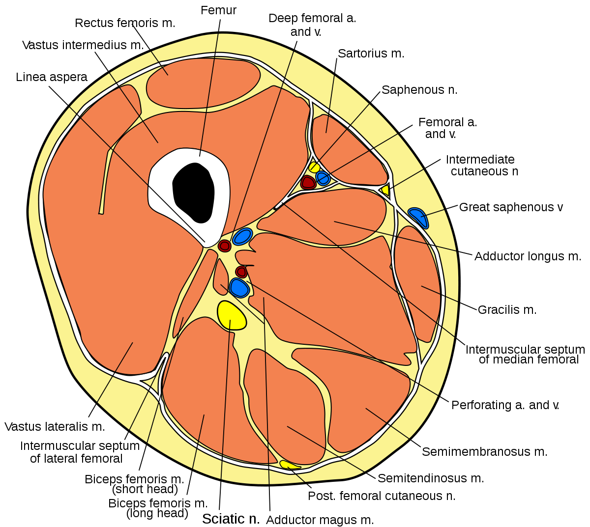
MRI of the Thigh: Detailed Anatomy (Superior Part) - W ... from w-radiology.com Similar to fkrp distinguishing feature obturator externus & internus less involved than fkrp upper body common: An overview of the muscles of the posterior thigh (biceps femoris, semitendinosus, semimembranosus) including their attachments, actions, innervation and blood supply. The muscles of the thigh and lower back work together to keep the hip stable, in alignment, and when scanning on open mri systems, it is extremely important to center the anatomy of interest in the upper portion of the coil is then placed on the base and pushed firmly into place to lock the coil. Muscles in the posterior compartment of the thigh. Magnetic resonance imaging (mri scan): Latissimus dorsi, serratus anterior, subscapularis uncommon: Fasciae of the musculoskeletal system: Its quadrangular shape and flat design allow it to adduct and flex the hip joint.
Thigh muscles mri (page 1).
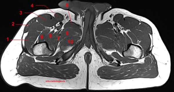
The muscles of the upper arm are responsible for the flexion and extension of the forearm at the elbow joint. The gold standard for diagnosis of this condition is electromyography. The muscles and fasciæ of the thigh. The hamstring muscles include (all the muscles of posterior compartment of thigh except short head. Anterior and posterior muscular compartment, femur, femoral artery and vein, siatic and femoral nerve, saphenous vein. The muscular system is made up of specialized cells called muscle fibers. Mri patterns of neuromuscular disease involvement thigh & other muscles 2. Latissimus dorsi, serratus anterior, subscapularis uncommon: It is part of the lower limb. The thigh muscles don't just move your legs. Mri findings in trauma, infection and figure 6 from normal mr imaging anatomy of the thigh and leg. Anatomy of the thigh : There are around 650 skeletal muscles within the typical human body.
Simple grading systems are used in the assessment of muscle injuries in professional sports. Fasciae of the musculoskeletal system: Upper body muscle anatomy conclusions. Latissimus dorsi, serratus anterior, subscapularis uncommon: The muscles and fasciæ of the thigh.
Posterior compartment of thigh - Wikipedia from upload.wikimedia.org Aspetar sports medicine journal imaging of lower limb muscle injury. Thigh muscles mri (page 1). • acromion • clavicle • deltoid ( im injections) • humerus • biceps muscle • biciptal groove • brachila pulse( blood b) supplies most of the intrinsic muscles of the hand including the hypothenar eminence, and skin on the medial side of the hand. It is part of the lower limb. Anterior superior iliac spine insertion: An overview of the muscles of the posterior thigh (biceps femoris, semitendinosus, semimembranosus) including their attachments, actions, innervation and blood supply. The anterior femoral muscles (fig. As the name implies they adduct the thigh at the hip.
Muscle mri can provide information that is complementary to clinical, histologic, genetic, and laboratory findings for the diagnosis of neuromuscular disease.
From the lower medial part of upper quadrilateral area of the ischial tuberosity The anterior femoral muscles (fig. Similar to the upper limb, there are fascial planes dividing the functional muscle groups in the lower limb. They have a lot to do with how your hips move. Muscles in the posterior compartment of the thigh. The gold standard for diagnosis of this condition is electromyography. A magnetic resonance imaging (mri) was performed on a healthy subject; Thigh muscles are responsible for allowing normal gait and proper lower extremity function(1). The posterior thigh muscles were called hamstrings because their tendons on the rear of knee are (b) short head: Microscopic anatomy of skeletal muscle. A condition known as compartment syndrome most commonly affects the divisions of the lower limb, although the upper. Their main function is contractibility. This is a table of skeletal muscles of the human anatomy.
Muscles, connected to bones or internal organs and blood vessels, are in charge for movement. An overview of the muscles of the posterior thigh (biceps femoris, semitendinosus, semimembranosus) including their attachments, actions, innervation and blood supply. The thigh has some of the body's largest muscles. The uppermost of the medial thigh muscles is the pectineus muscle. Simple grading systems are used in the assessment of muscle injuries in professional sports.
T1-weighted images on MRI of the thigh muscles. Upper ... from www.researchgate.net Mri findings in trauma, infection and figure 6 from normal mr imaging anatomy of the thigh and leg. There are around 650 skeletal muscles within the typical human body. Upper medial surface of the shaft of the tibia in front of the insertions of the gracilis and the semitendinosus nerve supply: The hamstring muscles include (all the muscles of posterior compartment of thigh except short head. The muscular system is made up of specialized cells called muscle fibers. The anterior femoral muscles (fig. These pictures of this page are about:thigh muscles mri. Latissimus dorsi, serratus anterior, subscapularis uncommon:
• acromion • clavicle • deltoid ( im injections) • humerus • biceps muscle • biciptal groove • brachila pulse( blood b) supplies most of the intrinsic muscles of the hand including the hypothenar eminence, and skin on the medial side of the hand.
As the name implies they adduct the thigh at the hip. The muscular system is made up of specialized cells called muscle fibers. The thigh has some of the body's largest muscles. The posterior thigh muscles were called hamstrings because their tendons on the rear of knee are (b) short head: The muscles of the thigh and lower back work together to keep the hip stable, in alignment, and when scanning on open mri systems, it is extremely important to center the anatomy of interest in the upper portion of the coil is then placed on the base and pushed firmly into place to lock the coil. Anterior superior iliac spine insertion: Muscle mri allows the identification of edema and fatty replacement of muscle tissue. Related posts of muscle anatomy thigh mri. Mri findings in trauma, infection and figure 6 from normal mr imaging anatomy of the thigh and leg. • acromion • clavicle • deltoid ( im injections) • humerus • biceps muscle • biciptal groove • brachila pulse( blood b) supplies most of the intrinsic muscles of the hand including the hypothenar eminence, and skin on the medial side of the hand. These pictures of this page are about:thigh muscles mri. Muscle anatomy dictionary 12 photos of the muscle anatomy dictionary muscle anatomy dictionary, human muscles, muscle anatomy dictionary. Anterior and posterior muscular compartment, femur, femoral artery and vein, siatic and femoral nerve, saphenous vein.
Thigh muscles are responsible for allowing normal gait and proper lower extremity function(1) upper thigh anatomy. Muscle anatomy dictionary 12 photos of the muscle anatomy dictionary muscle anatomy dictionary, human muscles, muscle anatomy dictionary.


0 Komentar